bio-medical tomography
Fig. 6.
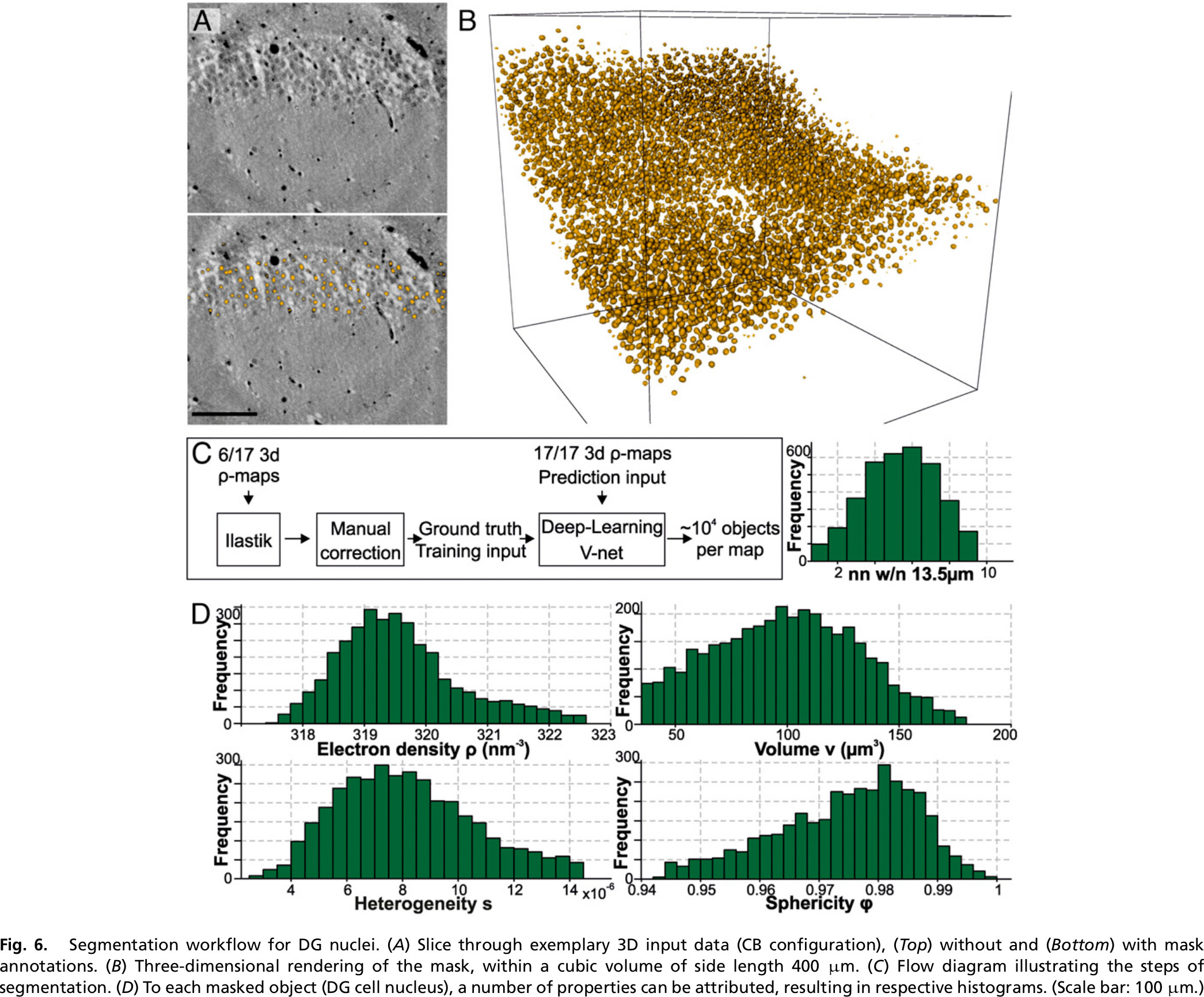
Segmentation workflow for DG nuclei.
(A) Slice through exemplary 3D input data (CB configuration), (Top) without and (Bottom) with mask annotations.
(B) Three-dimensional rendering of the mask, within a cubic volume of side length 400 μm.
(C) Flow diagram illustrating the steps of segmentation.
(D) To each masked object (DG cell nucleus), a number of properties can be attributed, resulting in respective histograms.
(Scale bar: 100 μm.)
Publications
Three-dimensional virtual histology of the human hippocampus based on phase-contrast computed tomography
Eckermann M, Schmitzer B, van der Meer F, Franz J, Hansen O, Stadelmann C, Salditt T - Proceedings of the National Academy of Sciences - 2021
IMAGED OBJECTS
segmented DG nuclei, human hippocampus
Bio-medical tomography in Helmholtz Imaging CONNECT:
No lab found.
No solution found.