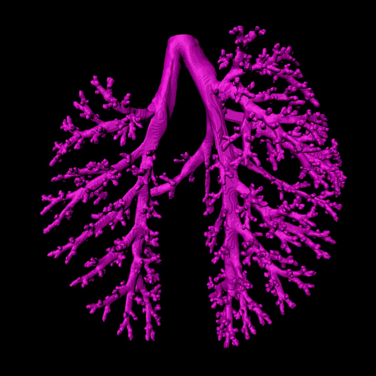
Covid-19 lung tissue visualisation
Study of the changes caused by the coronavirus in the structure of alveoli (the tiny air sacs in the lung) and the vasculature. They use multi-scale phase contrast x-ray tomography extending conventional histology by a third dimension and allowing for full quantification of tissue remodeling.
Resources used
DESY News: PETRA III provides new insights into Covid-19 lung tissue. In DESY, Deutsches Elektronen-Synchrotron. Retrieved 17:28, February 4, 2022, from https://www.desy.de/news/news_search/index_eng.html?openDirectAnchor=1894&two_columns=0
Publications
3D virtual pathohistology of lung tissue from Covid-19 patients based on phase contrast X-ray tomography
Eckermann M, Frohn J, Reichardt M, Osterhoff M, Sprung M, Westermeier F, Tzankov A, Werlein C, Kühnel M, Jonigk D, Salditt T - eLife - 2020