X-ray imaging (XRI)
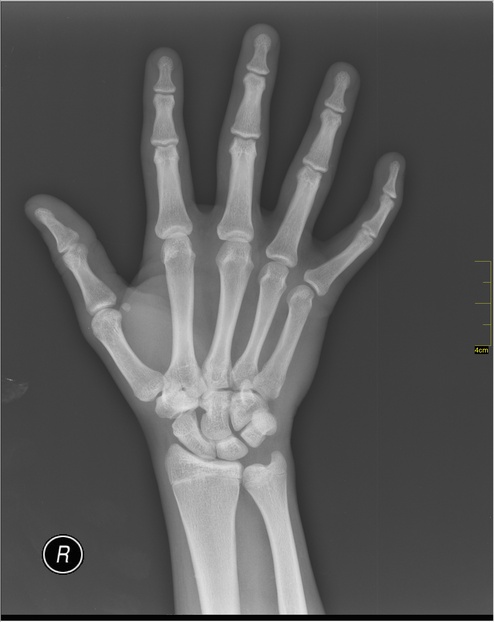
X-ray imaging is an imaging technique where a target's inner body is observed by measuring the amount of energy loss when irradiating the target with X-rays. Those rays are electromagnetic waves in the spectrum 1 nm–1 pm, or (30 PHz–30 EHz), and carry more energy than visible light (430 THz–750 THz), or ultraviolet (300 THz–30 PHz), as photon energy (in eV) is proportional to its frequency. It is in particular used in medical imaging, as they interact with human organs in different ways. Soft tissues like muscles absorb little X-ray energy and therefore are rendered on an X-ray image as high intensities, creating contrast in the images.
Resources used
X-ray. In Wikipedia, The Free Encyclopedia. Retrieved 17:28, February 4, 2022, from https://en.wikipedia.org/wiki/X-ray
See also:
Experimental and Computational Methods at Institute of Resource Ecology