Precession Electron Diffraction & Strain Mapping (NANOMEGAS) (Precession Electron Diffraction)
Precession electron diffraction
Precession electron diffraction (PED) is a specialized method to collect electron diffraction patterns in a transmission electron microscope (TEM). By rotating (precessing) a tilted incident electron beam around the central axis of the microscope, a PED pattern is formed by integration over a collection of diffraction conditions. This produces a quasi-kinematical diffraction pattern that is more suitable as input into direct methods algorithms to determine the crystal structure of the sample.
Read more about 'Precession electron diffraction' at: WikipediaWikipedia contributors. "Precession electron diffraction." Wikipedia, The Free Encyclopedia. Wikipedia, The Free Encyclopedia, Feb. 29, 2024.
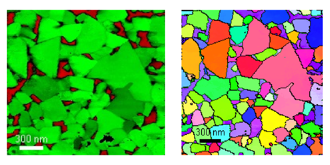
Precession Electron Diffraction is a transmission electron microscopy technique in which a condensed electron beam is tilted off axis and rotated, recording the electron diffraction pattern. The images collected are integrated over the whole rotation, giving a much wider view of the diffraction conditions. The spot is then rastered over the sample allowing a map of the change in diffraction conditions to be made. From this we can look at the Phase and Orientation of the sample, or perform Strain Mapping.