TFS Spectra 300 (Spectra)
Jülich
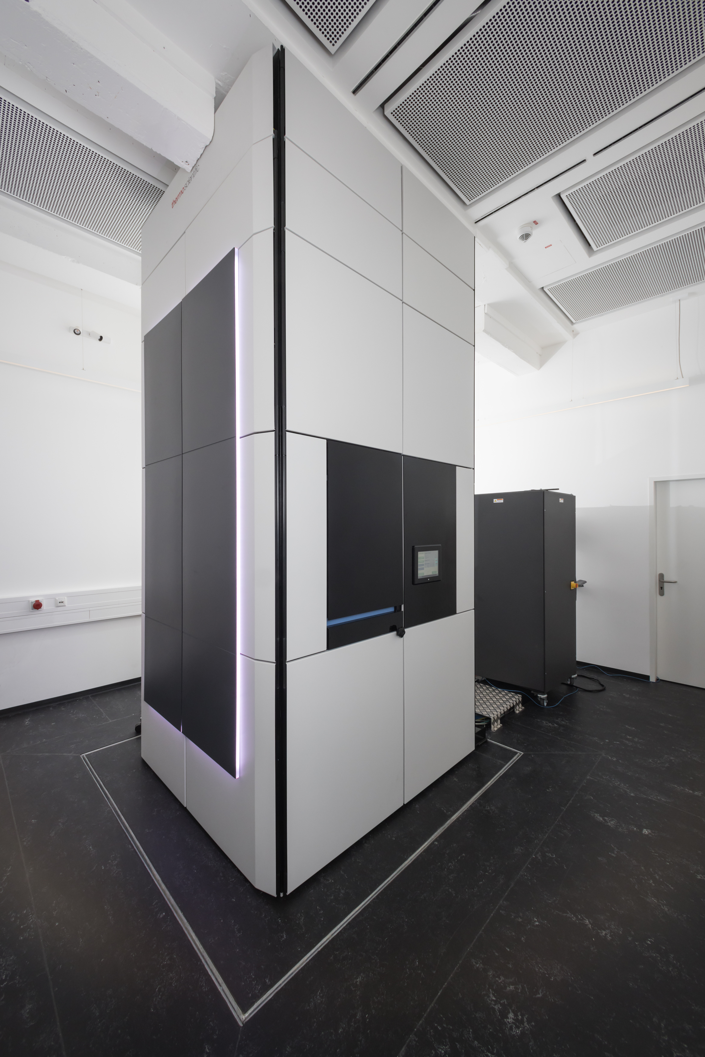
The TFS Spectra 300 is a state-of-the-art FEG Scanning Transmission Electron Microscope (S/TEM) with a high-tension voltage range of 30 kV – 300 kV. It is completely enclosed in a casing, specially designed to dampen acoustic and temperature variations from the environment. This enclosure not only makes it possible to transfer information well below 1 Å resolution, it also allows the system to easily reach ultra-high resolution routinely in a noisier environment. The Spectra 300 has been designed for the investigation of a wide range of solid-state phenomena taking place on the atomic scale of both the structure and chemical composition. For these purposes, the instrument is equipped with a high-brightness X-Feg monochromated source, a piezo-enhanced CompuStage, and two Cs correction optics. The S-CORR above the (S-TWIN) objective lens is used to form electrons probes with sub-Ångström. The CETCOR below the objective lens can be used for high-resolution TEM imaging with a bottom-mounted, retractable, fast Ceta CMOS camera. A variety of detectors are available, such as a Super-X detector with effectively 0.7 srad collection angle, a Gatan Continuum 1066 energy filter (GIF), a multichannel segmented Panther detector, a pixelated EMPAD detector, bright-field and dark-field detectors of the GIF, and a standard Fischione dark-field detector. Optical alignment are available for the beam energies 30 keV, 60 keV, 200 keV, and 300 keV.
Sample Environment
The TFS Spectra 300 allows samples to be investigated under room temperature at a vacuum level of about 10–8 mbar. Besides this standard setup, the sample environment can be adapted to various conditions, e.g. the thermal treatment or the application of external electric or magnetic fields to samples, making use of a wide portfolio of in situ TEM holders available through the ER-C user services.
Typical Applications and Limitations of Use
The multitude of detectors enables the application of a wide variety of microscopy techniques with focus on investigations at high spatial resolution. Among STEM based techniques, the instrument offers elemental mapping using energy dispersive X-ray (EDX) and electron energy loss (EEL) spectroscopy in STEM, as well as differential phase contrast (DPC) and scanned diffraction imaging (4D-STEM). Other available techniques are electron tomography for TEM and STEM, STEM micro-diffraction, and Cs-corrected field-free Lorentz imaging. Depending on detector configurations, some of the techniques can be applied in parallel for multi-modal imaging.
Link: https://er-c.org/index.php/access/